Abstract Authors
Michaela Davies - Rhodes University Biotechnology Innovation Centre (RUBIC), Department of Biotechnology, Rhodes University
Ronen Fogel - Rhodes University Biotechnology Innovation Centre (RUBIC), Department of Biotechnology, Rhodes University
Janice Limson - Rhodes University Biotechnology Innovation Centre (RUBIC), Department of Biotechnology, Rhodes University
Abstract Description
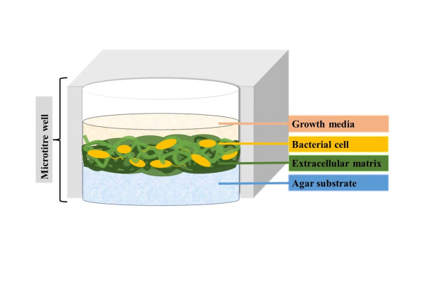
Biofilms are complex, protective communities of bacterial cells that adhere both to a substrate and to one-another through the secretion of extracellular matrices. Biofilm-associated cells often exhibit extremely different phenotypic and physiological characteristics compared to their dispersed/planktonic counterparts, most notably their enhanced resistance against antimicrobial agents. Consequently, biofilm-associated cells represent a more relevant model compared to dispersed cells for drug screening applications. Numerous fluorescent and colorimetric stains have reportedly been used to monitor biofilm formation and growth, however; a systematic comparison reviewing these stains has not yet been conducted. In this study, the ability of established colorimetric and fluorometric stains in detecting distinct biofilm components - compared to dispersed/planktonic cells - was evaluated, using Escherichia coli as a model organism. The stains were assessed based on two key parameters: specificity – the ability to differentiate biomass signals from negative controls (with particular consideration of interference from growth media), and sensitivity – the ability to produce a statistically-significant increase in signal corresponding to increasing cell density in both dispersed and biofilm-associated cell samples.