Abstract Authors
Mfundo Magagula - Department of Biochemistry, Microbiology and Biotechnology, University of Limpopo
Thabiso Motaung - Department of Biochemistry, Microbiology and Biotechnology, University of Limpopo
Zukile Mbita - Department of Biochemistry, Microbiology and Biotechnology, University of Limpopo
Khumisho Dithebe - Department of Biochemistry, Microbiology and Biotechnology, University of Limpopo
Abstract Description
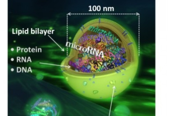
Fungi produce a variety of extracellular vesicles (EVs) that transport distinct cargoes to facilitate intercellular communication and influence the physiology of the recipient cells. Fungal endophytes may use EV-enclosed secondary metabolites and enzymes to initiate host plant colonisation and fend off invading pathogens. However, the bioactive and enzymatic potential of EVs of fungal endophytes remain underexplored. The current study characterised and screened the antimicrobial and enzymatic capabilities of EVs isolated from the endophytic fungi Neofusicoccum parvum KaS-3 and Pseudofusicoccum olivaceum KaS-5. Size exclusion chromatography was used to isolate EVs from cell-free fermented potato dextrose broth after 3 days, while ethyl acetate was used to extract secondary metabolites following a 21-day incubation period. The size and concentration of the EVs were assessed using Nanoparticle Tracking Analysis. Antimicrobial activity of the EVs and crude extracts were assessed using broth microdilution assay against Escherichia coli, Staphylococcus aureus and Candida albicans. An agar-based susceptibility assay was also performed to confirm antimicrobial activity. Amylase, laccase, lipase and protease activities of the isolates and their respective EVs were screened using the agar plate assay, while peroxidase activity was evaluated using the test tube method. The sizes of the EVs ranged between 75–395 nm and particle concentrations were 2.34 x 10⁹ and 1.23 x 10⁹ for N. parvum KaS-3 and P. olivaceum KaS-5, respectively. Antimicrobial screening revealed that only the crude extract of N. parvum KaS-3 exhibited inhibitory activity against E. coli, S. aureus and C. albicans, with minimum inhibitory concentration (MIC) values of 1.25, 2.5 and 2.5 mg/mL, respectively. In contrast, the crude extract of P. olivaceum KaS-5 and EVs from both isolates showed no activity against the tested pathogens. Enzyme screening revealed that both the isolates produce all the screened enzymes, except laccase, while the EVs only exhibited protease and lipase activities. The study highlights the potential of fungal endophyte EVs as alternative sources of hydrolytic enzymes with applications in various biotechnological industries.